Serial Two-Photon Tomography Whole-Tissue Imaging System
Serial Two-Photon Tomography Whole-Tissue Imaging System
|
| description: |
The Automated Serial Two-Photon 3D Whole-Tissue Imaging System is an automated research platform designed for three-dimensional imaging and spatial analysis of intact mouse brains and large-volume tissue specimens. The system uses a precision microtome to |
Product Overview
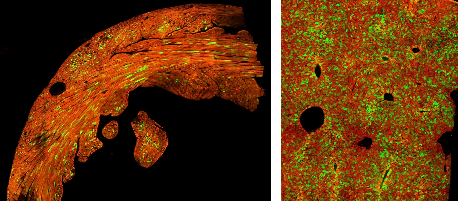
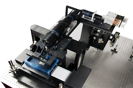
The Automated Serial Two-Photon 3D Whole-Tissue Imaging System is an automated research platform designed for three-dimensional imaging and spatial analysis of intact mouse brains and large-volume tissue specimens. The system uses a precision microtome to serially section fixed tissue and performs two-photon fluorescence imaging immediately after each section is generated. Hundreds or thousands of sequential images can then be automatically registered, stitched, and reconstructed into a high-resolution 3D dataset, providing comprehensive spatial information ranging from individual cells and neural fibers to large-scale brain structures.
Unlike conventional two-photon microscopes, which typically focus on localized regions of tissue, the system is designed to image entire brains or large tissue volumes as integrated 3D specimens. Key applications include neuronal and neural-circuit tracing, brain-region connectivity analysis, vascular mapping, neurodegenerative disease research such as Alzheimer's disease, and whole-brain biodistribution studies for AAV and gene therapies. The serial tissue sections can also be preserved for immunohistochemistry, histology, and molecular analysis, allowing these conventional tissue-based results to be correlated with their original three-dimensional spatial locations.
Product Advantages
Whole-Brain 3D Imaging: Enables high-resolution 3D imaging of intact mouse brains and large tissue volumes.
Automated Serial Sectioning: Integrates automated sectioning and two-photon imaging in a continuous section–image workflow.
High-Resolution Spatial Information: Reveals 3D spatial relationships among cells, neural fibers, blood vessels, and brain structures.
Tissue Preservation: Serial sections can be retained for subsequent immunohistochemistry and histological analysis.
Whole-Brain Applications: Particularly suitable for neural circuits, brain diseases, and AAV/gene therapy biodistribution studies.
Main Product Features
Multichannel Fluorescence Imaging: Enables detection and analysis of multiple fluorescent labels.
Precision Microtome: Uses a dedicated high-precision sectioning mechanism for stable and uniform serial tissue sectioning.
Automated Image Acquisition: Integrates sectioning and imaging for continuous and unattended data collection.
Large-Scale Data Processing: Supports registration, stitching, and 3D reconstruction of large volumes of sequential images.
Flexible Sample Handling: Suitable for fixed tissue samples of different sizes and types.
3D Visualization and Analysis: Enables three-dimensional visualization, localization, and quantitative analysis of reconstructed tissue datasets.
 Application Areas
Polymer Materials: Investigation of rapid crystallization, melting, glass transitions, and the effects of thermal history on material properties.
Metal Materials: Study of melting, crystallization, and phase-transition behavior under rapid heating and cooling conditions.
3D Printing Materials: Real-time investigation of laser-induced heating and cooling of 3D-printing powders.
Nanomaterials: Characterization of thermal properties and stability of thin films, nanoparticles, and other microscale materials.
Pharmaceutical Materials: Investigation of thermal behavior, crystallization kinetics, and the influence of processing conditions on pharmaceutical materials.
Crystallization Kinetics: Study of crystal formation and growth rates under different heating and cooling conditions.
Phase Transitions: Investigation of material-state changes, such as transitions from solid to liquid.
Material Thermal Stability: Analysis of material degradation and decomposition behavior at elevated temperatures.
Application Areas
Polymer Materials: Investigation of rapid crystallization, melting, glass transitions, and the effects of thermal history on material properties.
Metal Materials: Study of melting, crystallization, and phase-transition behavior under rapid heating and cooling conditions.
3D Printing Materials: Real-time investigation of laser-induced heating and cooling of 3D-printing powders.
Nanomaterials: Characterization of thermal properties and stability of thin films, nanoparticles, and other microscale materials.
Pharmaceutical Materials: Investigation of thermal behavior, crystallization kinetics, and the influence of processing conditions on pharmaceutical materials.
Crystallization Kinetics: Study of crystal formation and growth rates under different heating and cooling conditions.
Phase Transitions: Investigation of material-state changes, such as transitions from solid to liquid.
Material Thermal Stability: Analysis of material degradation and decomposition behavior at elevated temperatures.
Detailed Specifications
Product Specifications
Product Type: Automated Serial Two-Photon 3D Whole-Tissue Imaging System
Imaging Technology: Serial Two-Photon Tomography Plus (STP+)
Imaging Modality: High-speed multi-foci two-photon fluorescence imaging
Sample Type: Fixed intact organs and large-volume tissue specimens, particularly tissues from small animal models such as mice and rats
Spatial Resolution: Sub-micron in-plane (XY) resolution; representative mouse-brain datasets can reach approximately 1.2 µm XY sampling
Z-Axis Sampling: Flexible coronal or sagittal section spacing according to experimental requirements
Fluorescence Imaging: Multichannel and multispectral fluorescence imaging
Sample Preparation: Simple fixation and agar embedding; no optical clearing required
Sectioning Technology: 4th-generation FlexureSlice precision microtome
Serial Sectioning: Automated continuous tissue sectioning and imaging
Section Preservation: Imaged tissue sections can be collected for subsequent IHC, histology, and spatial biology analysis
Automated Slide Mounting: The TissueCyte 1600FC integrated section-capture system supports a standard capacity of 300 glass slides
3D Reconstruction: Sequential images are registered, stitched, and reconstructed into complete 3D tissue datasets
Data Output: High-resolution multichannel 2D images and volumetric 3D datasets
Imaging Scale: From tissue regions to complete organs
Operation: Designed for continuous automated operation and multi-sample studies
Imaging Speed: Whole-tissue imaging can be completed on a timescale of hours, depending on tissue size, sampling interval, and imaging settings.


 Scan QR Code & Subscribe